lunes, 26 de julio de 2010

INTRODUCTION


If you like good music just put play here, but if you want to watch the videos then you have to stop the music:


Currently the major advances in scientific research techniques have been settled much of the unknowns that for a long time, have remained unanswered in the fields of genetics. Putting in evidence a project that has been trying to get out into the open but this has a drawback that goes against moral ethics and calls itself “The Human Genome Project” Whose purpose is through genetics to develop a list of possible diseases, syndromes or complex that the human being is likely to have or already developed, on the other side are the benefits are prevent and cure diseases.

One of the most important developments is the discovery of the double helix structure of DNA, made in 1953 by Watson and Crick biologist, a discovery that caused the foundations of modern molecular biology. Actually this is a discovery of great importance to a lot of new processes. The contents of this information have been shown to depend on the order in which they have different types of nucleic acids to form DNA strands. This sequence is read the same way as you read various letters of the alphabet that make up a word, and are interpreted as a set of rules apply to all living beings and recently discovered which are called the human genome or genetic code. Other significant progress made in the field of genetics is the discovery of mutation sands their influence on living beings.

TOPIC 1


1.   STRUCTURE AND COMPOSITION OF DNA

•     CHOROMOSOME STRUCTURE:

In the nucleous of each cell, the DNA molecule is packaged into thread like structures called chromosomes. Each chromosome is made up of DNA tightly coiled many times around proteins called histones that support its structure. They are not visible even under a microscope.

Each chromosome has a constriction point called centromere, which divides the chromosome into two sections, or arms. The short arm of the chromosome is labeled the p arm. The long arm of the chromosome is labeled the q arm. The location of the centromere on each chromosome gives the chromosome its characteristic shape, and can be used to help describe the location of specific genes.

Almost all the analyses of the chromosomes are during the mitotic metaphase. During that phase of the cell cycle, DNA has been replicates and the chromatin is highly condensed. The Centromere is the structure where the mitotic spindle attaches prior to segregation.

The centromere can be established anywhere, if it is in the centre it is called metacentric, if it is a little bit up in the center it is called submetacentric, if it is close to the end of each chromosome it is called acrocentric and finally if it is at the end it is called telocentric.

Talking about this theme it would be better to understand for you if you can watch this video. This video was made by Dr. Stephen Sullivan.


What is DNA?


Deoxyribonucleic acid is a nucleic acid that contains the genetic instructions used in the development and functioning of all known living organisms and some viruses. The main role of DNA molecules is the long-term storage of information. DNA is often compared to a set of blueprints, like a recipe or a code, since it contains the instructions needed to construct other components of cells, such as proteins and RNA molecules.

The DNA segments that carry this genetic information are called genes, but other DNA sequences have structural purposes, or are involved in regulating the use of this genetic information.

The information in DNA is stored as a code made up of four chemical bases: adenine (A), guanine (G), cytosine (C), and thymine (T). Human DNA consists of about 3 billion bases, and more than 99 percent of those bases are the same in all people. The order, or sequence, of these bases determines the information available for building and maintaining an organism, similar to the way in which letters of the alphabet appear in a certain order to form words and sentences.


DNA bases pair up with each other, A with T and C with G, to form units called base pairs. Each base is also attached to a sugar molecule and a phosphate molecule. Together, a base, sugar, and phosphate are called a nucleotide. Nucleotides are arranged in two long strands that form a spiral called a double helix. The structure of the double helix is somewhat like a ladder, with the base pairs forming the ladder’s rungs and the sugar and phosphate molecules forming the vertical sidepieces of the ladder.

An important property of DNA is that it can replicate, or make copies of itself. Each strand of DNA in the double helix can serve as a pattern for duplicating the sequence of bases. This is critical when cells divide because each new cell needs to have an exact copy of the DNA present in the old cell.

Knowing what DNA is composed of is only half of the mystery, as scientists still could not work out the physical structure of the molecule.


What is a nucleotide?


Nucleotides are molecules that, when joined together, make up the structural units of RNA and DNA. In addition, nucleotides play central roles in metabolism. In that capacity, they serve as sources of chemical energy (adenosine triphosphate and guanosine triphosphate), participate in cellular signaling (cyclic guanosine monophosphate and cyclic adenosine monophosphate), and are incorporated into important cofactors of enzymatic reactions (coenzyme A, flavin adenine dinucleotide, flavin mononucleotide, and nicotinamide adenine dinucleotide phosphate).


What is a gene?

A gene is a unit of heredity in a living organism. It is normally a stretch of DNA that codes for a type of protein or for an RNA chain that has a function in the organism. All living things depend on genes, as they specify all proteins and functional RNA chains. Genes hold the information to build and maintain an organism's cells and pass genetic traits to offspring. A modern working definition of a gene is "a locatable region of genomic sequence, corresponding to a unit of inheritance, which is associated with regulatory regions, transcribed regions, and or other functional sequence regions ".

- Structure:

There are two general types of gene in the human genome: non-coding RNA genes and protein-coding genes.
Non-coding RNA genes represent 2-5 per cent of the total and encode functional RNA molecules. Many of these RNA's are involved in the control of gene expression, particularly protein synthesis. They have no overall conserved structure.
Protein-coding genes represent the majority of the total and are expressed in two stages: transcription and translation (see Gene expression). They show incredible diversity in size and organisation and have no typical structure. There are, however, several conserved features.

-Composition:

Due to the chemical composition of the pentose residues of the bases, DNA strands have directionality. One end of a DNA polymer contains an exposed hydroxyl group on the deoxyribose; this is known as the 3' end of the molecule. The other end contains an exposed phosphate group; this is the 5' end. The directionality of DNA is vitally important to many cellular processes, since double helices are necessarily directional (a strand running 5'-3' pairs with a complementary strand running 3'-5'), and processes such as DNA replication occur in only one direction. All nucleic acid synthesis in a cell occurs in the 5'-3' direction, because new monomers are added via a dehydration reaction that uses the exposed 3' hydroxyl as a nucleophile.

QUESTION PROBLEM TOPIC 1


a)Deduce the type of genetic material used  by:
·      Cattle
·      E.coli
·      Influenza viruses
b)Suggest a reason for the difference between Cattle thymus gland, Spleen and sperm in the measurements of their base composition.
c) -Explain the reasons for the total amount of adenine plus guanine being close to 50% in the genetic material of many of the species in the table.
-Identify two other trends in the base composition of the species that have 50% adenine and guanine.
d) -Identify a species shown in the table that does not follow the trends in base composition described in C)
-Explain the reasons for the base composition of this species being different.


Answers:


a)-Cattle: The type of material genetic by the cattle is used is DNA because it isn't using Uracil.
-E. Coli: The type of genetic material used by the E.Coli is DNA because it isn't using uracil it is using Thymine.
-Influenza Viruses: The type of the genetic material used by the influenza Viruses is RNA because it isn't using thymine it is using uracil.
b)We have different body parts in this case talking about the cattle parts, in the table apears different parts, maybe in the table are diffent kind of cattle that is the principal reason I think, or maybe there is somebody trying to do a test with that animal.
c)-In most of the situations in the table we have percentages close to 50 % sometimes more, sometimes less and sometimes the same thing. In DNA the adenine goes with the thymine, and the guanine with the cytosine, so a base might be adenine or guanine, so operating those bases the answer might be close to 50%.
-The answer is the wheat and the yeast.
yeast:31.3+18.7=50%
Wheat:27.3+ 22.7=50%
d)-The cattle thymus gland, The cattle sleen, the cattle sperm, the pig thymus gland, the salmon, E.coli, the human sperm and finally the influenza virus.
-Because it depends on the animal, the enviroment and that staff, so all species are different and the influenza is a virus so it use RNA.

TOPIC 2

2. Karyotypes:


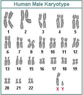
Karyotypes are images of chromosomes to display their banding patterns. When a nucleus is in during metaphase of mitosis, its chromosomes are condensed and the banding of the chromosomes can be visualized when certain dyes (e.g. Giemsa dye) are added to the chromosomes. There are several classical methods available to visualize the banding pattern as well as a more genomic one called chromosomal painting.

For those of us who are unaccustomed to seeing real chromosomes, often they are drawn in a cartoon fashion called an ideogram. Below is an ideogram of the X chromosome. The short arm of any chromosome is called the "p" arm which stands for the French word for small - petite. The long arm is called the "q" arm. Many years ago, histologist numbered the bands for each arm so we can refer to particular bands as genomic locations and everyone will be looking at the same band.


A karyotype is the number and appearance of chromosomes in the nucleous of a eukaryote cell. The term is also used for the complete set of chromosomes in a species, or an individual organism.


Karyotypes describe the number of chromosomes, and what they look like under a light microscope. Attention is paid to their length, the position of the centromeres, any differences between the sex chromosomes, and any other physical characteristics. The preparation and study of karyotypes is part of cytogenetics.

The study of whole sets of chromosomes is sometimes known as karyology. The chromosomes are depicted (by rearranging a microphotograph) in a standard format known as a karyogram or idiogram: in pairs, ordered by size and position of centromere for chromosomes of the same size.

Karyotypes can be used for many purposes; such as, to study chromosomal aberrations,cellular function, taxonomic relationships, and to gather information about past evolutionary events.


The karyotype is characteristic of each species, as the number of chromosomes, humans have 46 chromosomes (23 pairs because we are diploid or 2n) nucleus of every cell, [1] organized into 22 autosomes and 1 pair peer sexual (male XY and female XX). However, some individuals have other karyotypes with added or missing sex chromosomes, including 47,XYY47,XXY47,XXX and 45,X. The other possibility, 45,Y, does not occur, as an embryo with only a Y chromosome is incapable of survival.


Staining: The study of karyotypes is possible due to staining. Usually a colorant is applied after they have been arrested during cell division by colchicine solution. For the human white blood cells are the most frequently used because they are easily induced to grow and divide in tissue culture. Sometimes the comments can be made when cells are not dividing. The sex of a newborn can be determined by observation of cells at the interface. Most species have a standard karyotype.


Classic Karyotipe: In the "classic" (depicted) karyotype, a dye, often Giemsa (G-banding), less frequently Quinacrine, is used to stain bands on the chromosomes. Giemsa is specific for the phosphate groups of DNA. Quinacrine binds to the adenine-thymine-rich regions. Each chromosome has a characteristic banding pattern that helps to identify them; both chromosomes in a pair will have the same banding pattern. 


Karyotypes are arranged with the short arm of the chromosome on top, and the long arm on the bottom. Some karyotypes call the short and long arms p and q, respectively. In addition, the differently stained regions and sub-regions are given numerical designations from proximal to distal on the chromosome arms. For example, Cri du chat syndrome involves a deletion on the short arm of chromosome 5. It is written as 46,XX,5p-. The critical region for this syndrome is deletion of 15.2, which is written as 46,XX, of 5.


Spectral Karyotype: Spectral karyotyping is a molecular cytogenetic technique used to simultaneously visualize all the pairs of chromosomes in an organism in different colors. Fluorescently labeled probes for each chromosome are made by labeling chromosome-specific DNA with different fluorophores. Because there are a limited number of spectrally-distinct fluorophores, a combinatorial labeling method is used to generate many different colors. Spectral differences generated by combinatorial labeling are captured and analyzed by using an interferometer attached to a fluorescence microscope. Image processing software then assigns a pseudo color to each spectrally different combination, allowing the visualization of the individually colored chromosomes.


This technique is used to identify structural chromosome aberrations in cancer cells and other disease conditions when Giemsa banding or other techniques are not accurate enough.

Virtual Karyotype: detects genomic copy number variations at a higher resolution level than conventional karyotyping or chromosome-based comparative genomic hybridization.

QUESTION PROBLEM TOPIC 2


1. Find one normal karyotype on the male sex, and one normal Karyotype on the female sex, and then find 4 atypical karyotypes.
Answer:
1. NORMAL KARYOTYPE:
-Female Karyotype:contain two X chromosomes and are denoted 46,XX; males have both an X and a Y chromosome denoted 46,XY. Any variation from the standard karyotype may lead to developmental abnormalities.


-Male Karyotype: Contain one X chromosome and one Y chromosome.

ATYPICAL KARYOTYPES:
-Turner Syndrome: Turner syndrome (TS) is a medical disorder that affects about 1 in every 2,500 girls. Although researchers don't know exactly what causes Turner syndrome, they do know that it's the result of a problem with a girl's chromosomes (pronounced: krow-muh-soamz).
Most girls are born with two X chromosomes, but girls with Turner syndrome are born with only one X chromosome or they are missing part of one X chromosome. The effects of the condition vary widely among girls with Turner syndrome. It all depends on how many of the body's cells are affected by the changes to the X chromosome.
Girls with Turner syndrome are usually short in height. Girls with Turner syndrome who aren't treated for short stature reach an average height of about 4 feet 7 inches (1.4 meters). The good news is that when Turner syndrome is diagnosed while a girl is still growing, she can be treated with growth hormones to help her grow taller.
In addition to growth problems, Turner syndrome prevents the ovaries from developing properly, which affects a girl's sexual development and the ability to have children. Because the ovaries are responsible for making the hormones that control breast growth and menstruation, most girls with Turner syndrome will not go through all of the changes associated with puberty unless they get treatment for the condition. Nearly all girls with Turner syndrome will be infertile, or unable to become pregnant on their own.


-Down syndrome: Down syndrome is a congenital condition caused by an extra chromosome. The presence of an extra number 21 chromosome causes the distinctive facial features, physical characteristics and the cognitive impairments seen in people with Down syndrome. While people with Down syndrome have some characteristics in common, it is very important to remember that each person with Down syndrome is an individual with strengths and weaknesses. Never make assumptions about a person’s abilities based on their diagnosis. Having three copies of the genetic material on chromosome 21 is what causes Down syndrome.

-Medical problems
Children with Down syndrome are at higher risk to develop a number of specific medical problems. While most people with Down syndrome do not have serious medical problems, it is good to be aware of potential complications so that appropriate medical treatment can be sought early before serious complications arise.

Almost all infants with Down syndrome have low muscle tone which is called hypotonia. This means that their muscles are somewhat weak and they appear floppy. While this isn’t a medical problem per se, it is important because muscle tone can affect a child with Down syndrome’s ability to learn and grow. Hypotonia cannot be cured but it generally improves over time.
Hypotonia can also lead to some of the orthopedic or bone problems such at atlantoaxial instability that some people with Down syndrome can have.


The majority of children with Down syndrome will have some type of vision problem such as nearsightedness, farsightedness, crossed-eyes and even blocked tear ducts. About 40% of babies with Down syndrome are born with heart defects which can range from mild to severe. Somewhere between 40-60% of babies with Down syndrome will have some form of hearing loss. Other problems seen less frequently include gastrointestinal defects, thyroid problems and very rarely leukemia.


-Mental Retardation
All individuals with Down syndrome have some degree of mental retardation. They learn more slowly and have difficulties with complex reasoning and judgement, but they do have the capacity to learn. It is impossible to predict the degree of mental retardation in an infant with Down syndrome at birth (just as it is impossible to predict the IQ of any infant at birth).
It is very important that infant and people with Down syndrome receive the support, guidance, education and appropriate treatments needed to maximize their potential and to allow them to live fulfilling lives.

· Trisomy 18: There are 23 pairs of human chromosomes. In Trisomy 18 (Edwards syndrome), there is an extra chromosome with the 18th pair.

Trisomy 18 occurs in 1 in 3,000 live births. Unfortunately, most babies with Trisomy 18 die before birth, so the actual incidence of the disorder may be higher. Trisomy 18 affects individuals of all ethnic backgrounds.


-Trisomy 18 severely affects all organ systems of the body. Symptoms may include:
· Nervous system and brain - mental retardation and delayed development, high muscle tone,seizures, and physical malformations such as brain defects
· Head and face - small head (microcephaly), small eyes, wide-set eyes, small lower jaw
· Heart - congenital heart defects such as ventricular septal defect
· Bones - severe growth retardation, clenched hands with 2nd and 5th fingers on top of the others, and other defects of the hands and feet
· Malformations of the digestive tract, the urinary tract, and genitals


-Diagnosis
 The physical appearance of the child at birth will suggest the diagnosis of Trisomy 18. However, most babies are diagnosed before birth by amniocentesis (genetic testing of the amniotic fluid). Ultrasounds of the heart and abdomen can detect abnormalities, as can x-rays of the skeleton. 

· X X X syndrome: XXX syndrome (also called Trisomy X or Triple X) is caused by the presence of an extra ‘X’ chromosome in every cell. Typically, a female has two X chromosomes in every cell of their body, so the extra ‘X’ is unusual. The extra ‘X’ chromosome is typically inherited from the mother, but is a random event—not caused by anything she did or could prevent. Trisomy X is often not diagnosed until later in life, if ever. The risk of having a second child with an extra chromosome is approximately 1%, until mom is older than 38 years of age, as it is thought that this random event becomes more common as a woman ages. Prenatal testing is available in future pregnancies.

-Treatment: XXX syndrome is diagnosed prenatally, through CVS or amniocentesis, or after the child is born by a blood test. These tests are all able to look at a person’s chromosomes (karyotype.) There is no way to remove the extra X chromosome. Treatment depends on what needs the child has. Girls with XXX syndrome may need to be seen by physical, developmental, occupational, or speech therapists if they have developmental or speech problems. Additionally, a pediatric psychologist or group therapy may be helpful if they have social troubles. Girls with Trisomy X are treated as any other child with a developmental or psychological concern would be treated.


· Klinefelter syndrome: also known as the XXY condition, is a term used to describe males who have an extra X chromosome in most of their cells. Instead of having the usual XY chromosome pattern that most males have, these men have an XXY pattern.


Klinefelter syndrome is named after Dr. Henry Klinefelter, who first described a group of symptoms found in some men with the extra X chromosome. Even though all men with Klinefelter syndrome have the extra X chromosome, not every XXY male has all of those symptoms.


Because not every male with an XXY pattern has all the symptoms of Klinefelter syndrome, it is common to use the term XXY male to describe these men, or XXY condition to describe the symptoms.


Scientists believe the XXY condition is one of the most common chromosome abnormalities in humans. About one of every 500 males has an extra X chromosome, but many don’t have any symptoms.


For more information on genes and chromosomes.


-Symptoms
Not all males with the condition have the same symptoms or to the same degree. Symptoms depend on how many XXY cells a man has, how much testosterone is in his body, and his age when the condition is diagnosed.
The XXY condition can affect three main areas of development:

Physical development: As babies, many XXY males have weak muscles and reduced strength. They may sit up, crawl, and walk later than other infants. After about age four, XXY males tend to be taller and may have less muscle control and coordination than other boys their age.

As XXY males enter puberty, they often don’t make as much testosterone as other boys. This can lead to a taller, less muscular body, less facial and body hair, and broader hips than other boys. As teens, XXY males may have larger breasts, weaker bones, and a lower energy level than other boys.

By adulthood, XXY males look similar to males without the condition, although they are often taller. They are also more likely than other men to have certain health problems, such as autoimmune disorders, breast cancer, vein diseases, osteoporosis, and tooth decay.

XXY males can have normal sex lives, but they usually make little or no sperm. Between 95 percent and 99 percent of XXY males are infertile because their bodies don’t make a lot of sperm.


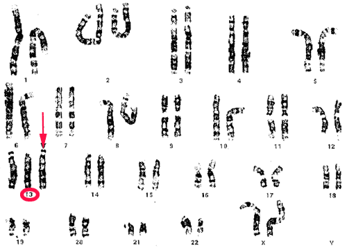
· Patau syndrome: Trisomy 13, is the least common of the autosomal trisomies, after Down syndrome (Trisomy 21) and Edwards syndrome (Trisomy 18). The extra copy of chromosome 13 in Patau syndrome causes severe neurological and heart defects which make it difficult for infants to survive. The exact incidence of Patau syndrome is not known, although it appears to affect females more than males, most likely because male fetuses do not survive until birth. Patau syndrome, like Down syndrome, is associated with increased age of the mother. It may affect individuals of all ethnic backgrounds.


-Symptoms
Newborns with Patau syndrome share common physical characteristics:
· Extra fingers or toes (polydactyly)
· Deformed feet, known as rocker-bottom feet
· Neurological problems such as small head (microcephaly), failure of the brain to divide into halves during gestation (holoprosencephaly), severe mental deficiency
· Facial defects such as small eyes (microphthalmia), absent or malformed nose, cleft lip and/or cleft palate
· Heart defects (80% of individuals)
· Kidney defects


-Treatment
Treatment of Patau syndrome focuses on the particular physical problems with which each child is born. Many infants have difficulty surviving the first few days or weeks due to severe neurological problems or complex heart defects. Surgery may be necessary to repair heart defects or cleft lip and cleft palate. Physical, occupational, and speech therapy will help individuals with Patau syndrome reach their full developmental potential.

TOPIC 3

4. Techniques of Analysis of Dna:


-DNA Fingerprinting:


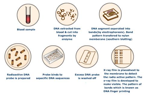
DNA fingerprinting is a way of identifying a specific individual, rather than simply identifying a species or some particular trait. It is also known as genetic fingerprinting or DNA profiling or DNA typing or DNA testing. DNA fingerprinting is currently used both for identifying paternity or maternity and for identifying criminals or victims. There is discussion of using DNAfingerprinting as a sort of personal identifier as well, although the viability of this is debatable.

The vast majority of a human's DNA will match exactly that of any other human, making distinguishing between two people rather difficult. DNA fingerprinting uses a specific type of DNA sequence, known as a microsatellite, to make identification much easier. Microsatellites are short pieces of DNA which repeat many times in a given person's DNA. In a given area, microsatellites tend to be highly variable, making them ideal for DNA fingerprinting. By comparing a number of microsatellites in a given area, one can identify a person relatively easily.

The sections of DNA used in DNA fingerprinting, although highly variable, are passed down from parents to their children. Although not all of the sections will necessarily be passed on, no child has pairs that their parents do not have. This means that by comparing large groups of these sections, paternity, maternity, or even both, may be determined. DNA fingerprinting has a high success rate and a very low false-positive rate, making it an extremely popular form of paternity and maternity verification.


-The Polymerase chain reaction (PCR):


The polymerase chain reaction (PCR) is a scientific technique in molecular biology to amplify a single or few copies of a piece of DNA across several orders of magnitude, generating thousands to millions of copies of a particular DNA sequence. The method relies on thermal cycling, consisting of cycles of repeated heating and cooling of the reaction for DNA melting and enzymatic replication of the DNA Primers (short DNA fragments) containing sequences complementary to the target region along with a DNA polymerase (after which the method is named) are key components to enable selective and repeated amplification. As PCR progresses, the DNA generated is itself used as a template for replication, setting in motion a chain reaction in which the DNA template is exponentially amplified. PCR can be extensively modified to perform a wide array of genetic manipulations.
Almost all PCR applications employ a heat-stable DNA polymerase, such as Taq polymerase, an enzyme originally isolated from the bacterium Thermus aquaticus. This DNA polymerase enzymatically assembles a new DNA strand from DNA building blocks, the nucleotides, by using single-stranded DNA as a template andDNA oligonucleotides (also called DNA primers), which are required for initiation of DNA synthesis. The vast majority of PCR methods use thermal cycling, alternately heating and cooling the PCR sample to a defined series of temperature steps. These thermal cycling steps are necessary first to physically separate the two strands in a DNA double helix at a high temperature in a process called DNA melting. At a lower temperature, each strand is then used as the template in DNA synthesis by the DNA polymerase to selectively amplify the target DNA. The selectivity of PCR results from the use of primers that are complementary to the DNA region targeted for amplification under specific thermal cycling conditions.


-Forensic Testing:
Forensic testing is the application of variou types of sciences in order to answer questions for the legal sytem. Testing takes various shapes and forms; one of the most common Modern approaches focuses on DNS testing.


There are several types of tests used by forensic scientists to identify both organic and inorganic substances involved in a crime. Fields involved include serology (blood testing), toxicology (the study of poisons) and DNA testing. Types of equipment used in forensic testing include spectrophotometers (to examine objects at wavelegths of light not visible to the naked eye), gas chromatographs (to separate components in a compound to analyse it more carefully) or fingerprinting analysis equipment (to compare a suspect's fingerprints with those found ata a crime scene).


-Paternity test:
A paternity test is conducted to prove paternity, that is, wheter a man is the biological father of another individual. This may be relevant in view of rights and duties of the father. Similarly, a maternity test can be carried out. 


-Restriction Enzymes:


Restriction enzymes are DNA-cutting enzymes found in bacteria (and harvested from them for use). Because they cut within the molecule, they are often called restriction endonucleases.

In order to be able to sequence DNA, it is first necessary to cut it into smaller fragments. Many DNA-digesting enzymes (like those in your pancreatic fluid) can do this, but most of them are no use for sequence work because they cut each molecule randomly. This produces a heterogeneous collection of fragments of varying sizes. What is needed is a way to cleave the DNA molecule at a few precisely-located sites so that a small set of homogeneous fragments are produced. The tools for this are the restriction endonucleases. The rarer the site it recognizes, the smaller the number of pieces produced by a given restriction endonuclease.

A restriction enzyme recognizes and cuts DNA only at a particular sequence of nucleotides.